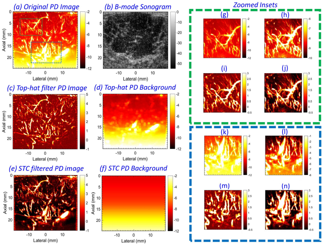
Figure 2:

Example of in vivo power Doppler (PD) (a) and B-mode (b) images from patient # 4. Background-suppressed PD image (c) and its corresponding noise field (d), obtained using top-hat based morphological filtering. Background-suppressed PD image (e) and its corresponding noise field (f), obtained using the proposed technique. For closer observation, the zoomed insets from green and blue regions of interest (ROIs) in (a) are displayed in (g-j) and (k-n), respectively. (g, k) display the PD data in (a). (h, l) display the PD data in (g, k), respectively, in a locally optimized color range to minimize the impact of the depth-dependent noise bias. (i, m) corresponds to zoomed insets from (c), and (j, n) correspond to zoomed insets from (e).
