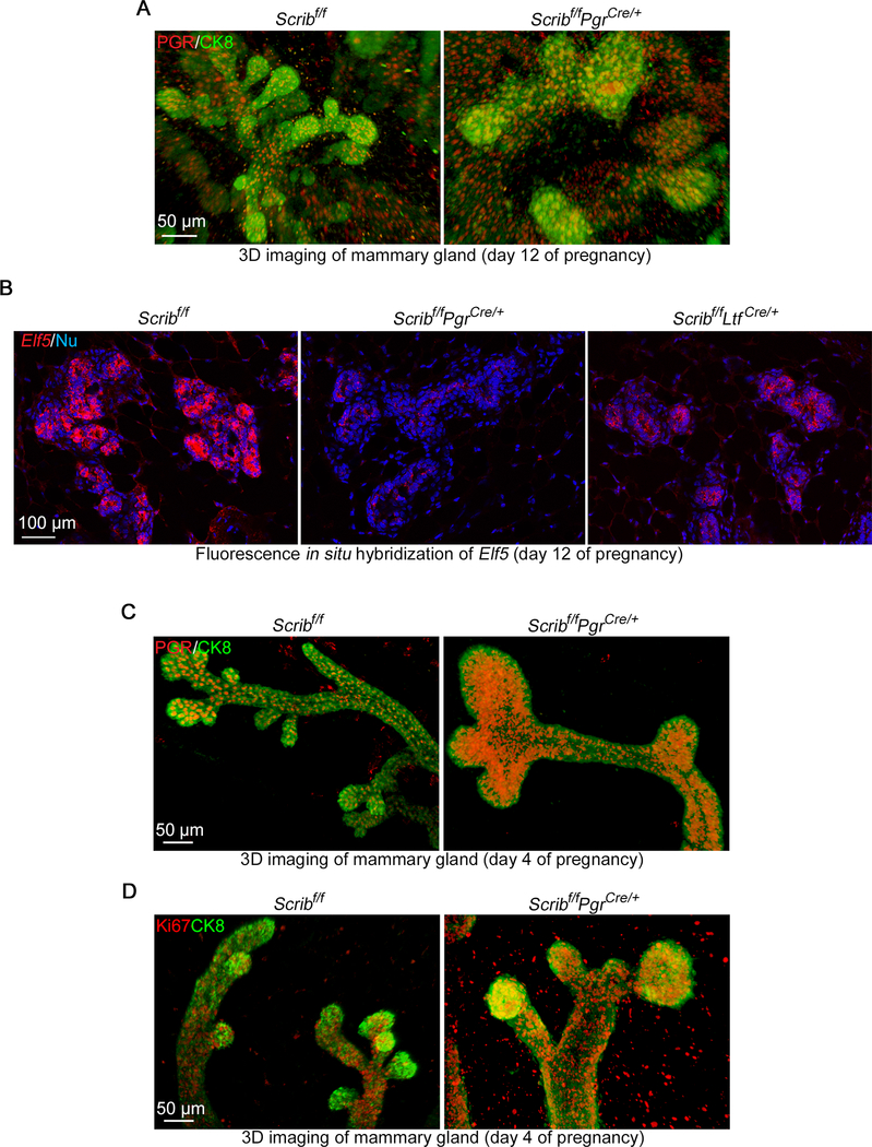
Figure 7. Scribf/fPgrCre/+ mammary glands fail to differentiate into mature alveolar cells.
A. Snapshots of tridimensional (3D) imaging of mammary glands from floxed and Scribf/fPgrCre/+ females on pregnancy day 12. Tissues were stained using antibodies for CK8 (epithelial marker) and PGR. Scale bar: 50 μm.
B. Fluorescence in situ hybridization of Elf5 in pregnancy day 12 mammary glands from females with each genotype. Scale bar: 100 μm.
C. Snapshots of 3D imaging of mammary glands from floxed and Scribf/fPgrCre/+ females on pregnancy day 4. Tissues were stained using antibodies for CK8 (epithelial marker) and PR. Scale bar: 50 μm.
D. Snapshots of 3D imaging of mammary glands from floxed and Scribf/fPgrCre/+ females on pregnancy day 4. Tissues were stained using antibodies for CK8 (epithelial marker) and Ki67. Scale bar: 50 μm.
Spotty signals outside of luminal layer in A, C and D are non-specific stainings caused by blood cells.
3D images were acquired by a Nikon multiphoton upright confocal microscope (Nikon A1R) using LWD 10X objective with 3 μm Z-stack.
Each image is a representative from at least 3 independent experiments.

