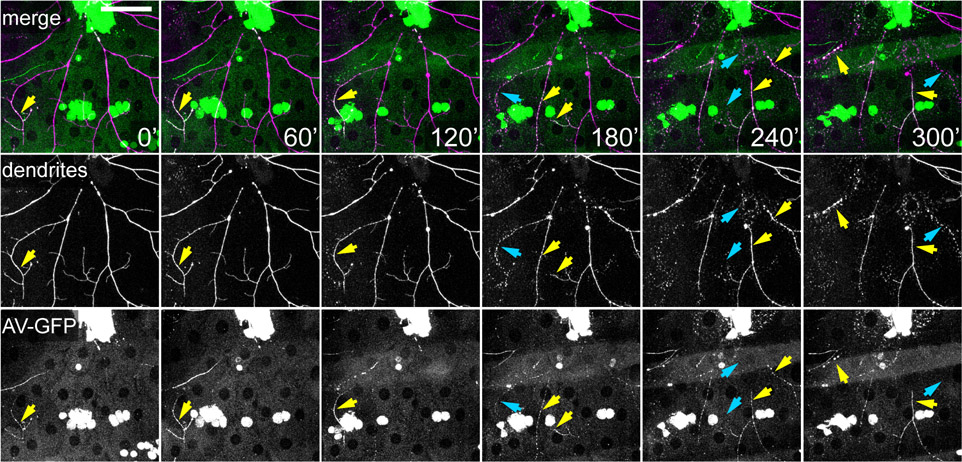
Figure 3: Time-lapse imaging of dendrite degeneration and exposure of an eat-me signal.
Selected frames from a time-lapse movie of degenerating dendrites of a class IV da neuron after laser injury. Dendrites were labeled by ppk>CD4-tdTom. The eat-me signal PS on degenerating dendrites was detected by Annexin-GFP (AV-GFP), which is secreted by the fat body. Yellow arrows point to the branches showing AV-GFP labeling. Blue arrows point to dendrites undergoing fragmentation.

