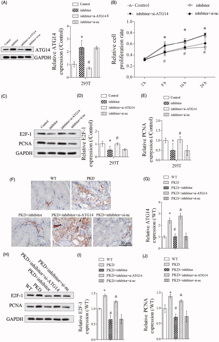
Figure 5.
ATG14 overexpression abolished the effect of miR-25-3p inhibitor on cell proliferation. PKD cells were divided into control, inhibitor, inhibitor + si-ATG14 and inhibitor + si-NC groups. (A) ATG14 expression was detected by Western blot. (B) Cell proliferation was detected by CCK-8 assay. (C–E) The expressions of E2F-1 and PCNA were detected by Western blot. N = 3. Mice were divided into WT, PKD, PKD + inhibitor, PKD + inhibitor + si-ATG14, PKD + inhibitor + si-NC groups. (F,G) The expression of ATG14 was analyzed by immunohistochemical technique. (H–J) The expressions of E2F-1 and PCNA were detected by Western blot. *p < 0.05 vs WT, #p < 0.05 vs PKD, &p < 0.05 vs PKD + inhibitor. N = 6.

