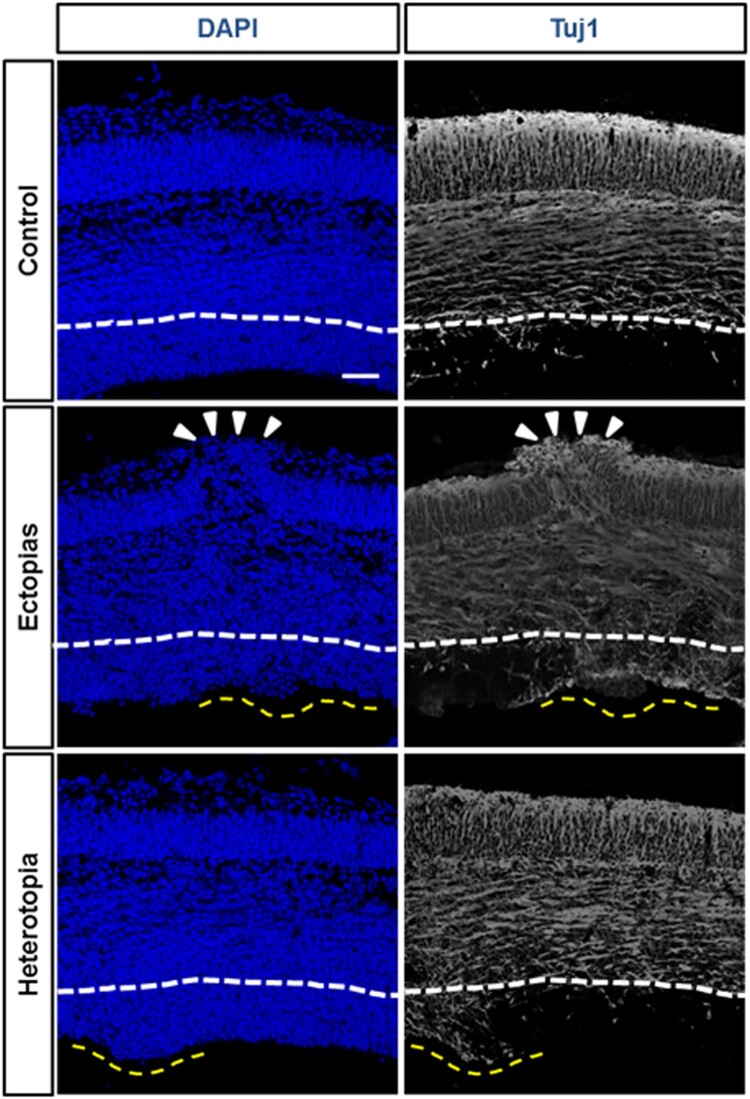
Figure 2.
Differentiation status of neurons at the ectopic and heterotopic region. The embryonic mouse brains at E13.5 stage were injected GFP-conjugated plasmids and harvested at E 15.5 stage. DAPI (blue) and Tuj1 (white) staining were performed to stain nucleus and differentiated neurons. The ectopia was marked white arrowheads and heterotopias were by a yellow dashed line. The white line was marked for the boundary layer of undifferentiated neuronal region as per the control set. The Tuj1-positive differentiated neuronal population was markedly increased in the ectopic and heterotopic cortical section. The scale bar was 50 μm.

