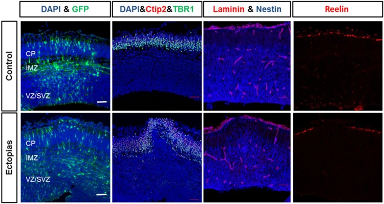
Figure 3.
Deformation of cortical layers in section cortex. The embryonic mouse brains at E13.5 stage were injected GFP-conjugated plasmids and harvested at E15.5 stage. (First panel) The neuronal migration was observed by GFP staining (green) in control and ectopic cortical sections. (Second panel) The transfected brain sections were stained with layer markers like Ctip2 (red) and TBR1 (green) and counterstained with the nuclear stain DAPI (blue). (Third panel) The transfected brain sections were stained with basal lamina marker laminin (red) and radial glial cell marker nestin (blue). (Fourth panel) The transfected brain sections were stained with telencephalon marginal zone marker Reelin (red). The scale bar was 50 μm.

