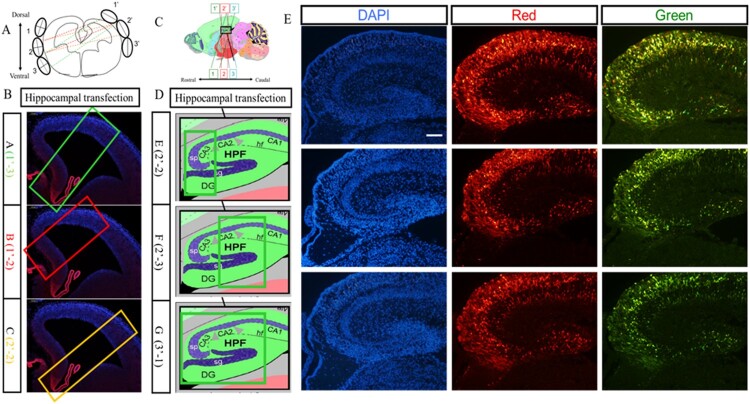
Figure 6.
Transfection in the hippocampus region of the embryonic brain. (A) Combinations of different positions of electrodes at the coronal plane for transfection. (B) Transfected regions at the coronal plane resulting from different combinations of electrodes. (C) Combinations of different positions of electrodes at the sagittal plane for transfection. (D) Transfected regions at the sagittal plane resulting from different combinations of electrodes. (E) The hippocampus transfected with RFP and GFP vectors simultaneously. Two kinds of plasmids with RFP and GFP were transferred into hippocampus at E14.5 stage by IUE, and red fluorescence protein and green fluorescence proteins were expressed in the hippocampus region. The scale bar was 100 μm.

