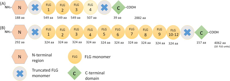
Figure 2.
Schematic representation of the proFilaggrin protein. (A) in dog and (B) in man. Note the differences in the number of filaggrin (FLG) repeats and in the number of the amino acids that form the FLG monomers and the C and N‐terminal domains (Brown and McLean 2012; Kanda et al. 2013; Pin et al. 2019). Abbreviations: aa, amino acid; FLG, filaggrin.

