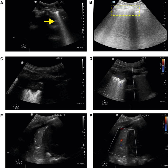
Fig. 3.
COVID-19 lung ultrasound findings. (A) B-lines (arrow); (B) irregular and broken pleural lines with multiple B-lines (dotted region); (C) peripheral or subpleural consolidation with (D) minimal colour Doppler signal. (E) Larger zone of consolidation in right lower posterior base with air bronchograms and (F) reduced perfusion using colour Doppler. (Courtesy of Dr. Stéphan Langevin and Dr. Caroline Gebhard) (Videos 3A, 3B, 3C, 3D, 3E, and 3F available as Electronic Supplementary Material)

