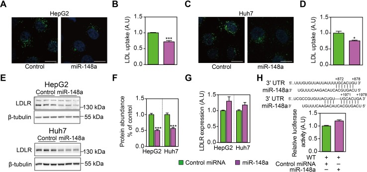
Fig 2. MiR-148a reduces cellular LDL uptake by reducing LDLR protein abundance but not its transcript levels.
HepG2 (A & B) and Huh7 cells (C & D) were transfected with control miRNA or miR-148a for 32h, and then treated with LSM for 16h before being incubated with 5 μg/mL Dylight-488-labeled LDL for 3h. A & C. Representative fluorescence images of cells. Nuclei are counterstained with 4’6-diaminido-2-phenylindole (blue). Scale bar: 10 μm. B & D. Quantitative measurement of LDL uptake in cells. Results are from four independent experiments in triplicates. N = 12; *: p<0.01; ***: p<0.001. E. HepG2 and Huh7 cells were transfected with control miRNA or miR-148a for 48 hours, and cultured under serum-containing medium. Total cell lysates were immunoblotted as indicated, and representative blots of 3 independent experiments in triplicates were shown. F. LDLR protein abundance was quantified and normalized to the level of tubulin in the same lysates, and expressed as the relative ratio of LDLR abundance in control miRNA transfected. N = 9. ***: p<0.001. G. HepG2 and Huh7 cells were transfected as above and culture under serum-containing medium, and LDLR mRNA levels were determined by quantitative PCR. Results are from four independent experiments in triplicates. N = 12; *: p<0.05. H. Putative miR-148a binding sites were identified in the 3’-UTR of human LDLR, and luciferase reporter plasmid was constructed accordingly. HEK293T cells were transfected the reporter plasmid, and cultured under serum-containing medium. Firefly luciferase activity was measured and corrected for Renilla luciferase activity in the same sample, and expressed as ratio of WT 3’-UTR transfected samples. Results are from four independent experiments in triplicates (N = 12).

