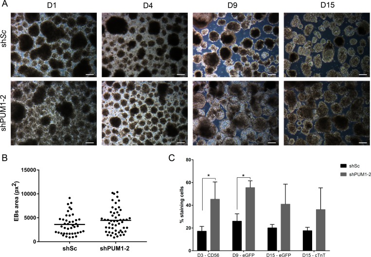
Fig 3. Effect of the knockdown of PUM1 and PUM2 during EB cardiac differentiation.
(A) Morphology of EBs at days 1 (D1), 4 (D4), 9 (D9) and 15 (D15) of the cardiomyogenic differentiation of hESCs previously transduced with shSc and shPUM1-2. Scale bars: 100 μm. (B) Area of EBs after 9 days of in vitro cardiac differentiation (the measurements were performed manually using ImageJ software) (n = 3). (C) Percentage of cells transduced with shSc and shPUM1-2 that were positive for CD56, eGFP/NKX2.5 and cTnT during cardiomyogenesis (n = 3). *p<0.05.

