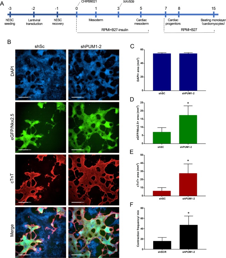
Fig 4. Effect of the knockdown of PUM1 and PUM2 during monolayer cardiac differentiation.
(A) Scheme of the protocol used for the transduction and monolayer cardiomyogenic differentiation of hESCs. (B) Representative immunofluorescence images of DAPI, eGFP/NKX2.5 and cTnT staining in the population of transduced hESCs after 15 days of monolayer cardiac differentiation. Scale bars: 500 μm. (C-E) The DAPI+ (C), eGFP/NKX2.5+ (D) and cTnT+ (E) stained areas were determined using Operetta CLS and Analysis Software 4.5 (Perkin Elmer) through a sequence analysis of 21 images (5X objective) obtained in triplicate after 15 days of monolayer cardiac differentiation (n = 3). (F) Number of contractions/minute during cardiac monolayer differentiation (n = 3). *p<0.05, **p<0.01.

