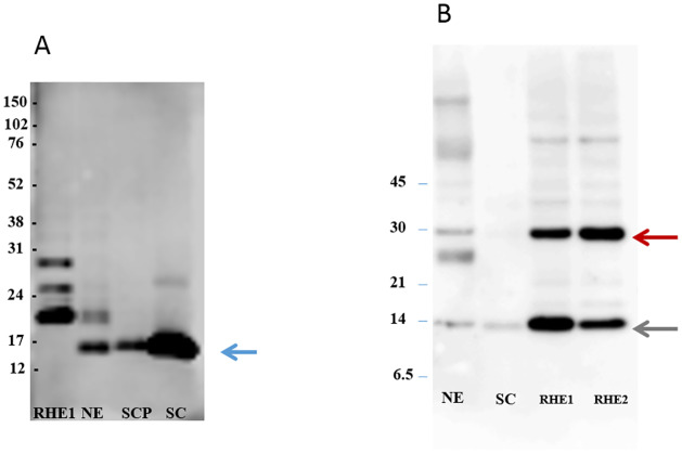
Fig 4. FLG2Nter is present in the stratum corneum of human skin in vivo.
A) Western blot analysis showing the detection of a 14 kDa band (blue arrow) representing the N-terminal domain of FLG2 in soluble protein extracts from the human stratum corneum and epidermis of normal skin. The contrast was enhanced from the original image (see supplementary information) to better visualize the bands. B) Western blot analysis showing the presence of SASPase 28 (red arrow) and the 14 kDa catalytic form of SASPase (green arrow) in epidermis of human skin and reconstructed skin. (RHE = reconstructed epidermal skin; NE = epidermis from normal skin; SCP = plantar stratum corneum; SC = stratum corneum (sampled by varnish stripping). Protein Molecular Weight markers are indicated on the left of each image.

