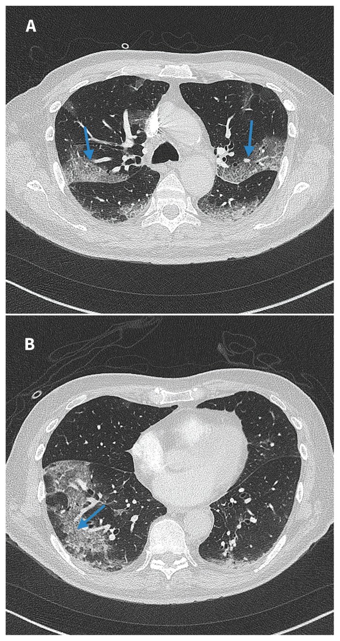
Figure 1:
(A) and (B) Computed tomography images of the chest (taken on day 3 of admission to hospital) of a 76-year-old man with coronavirus disease 2019 (COVID-19) and negative results for nasopharyngeal swabs. Bilateral peripheral ground glass opacification with areas of visible septal lines constituting crazy-paving are visible (blue arrows). This is typical of COVID-19 appearance as per the Radiological Society of North America Expert Consensus Statement.1

