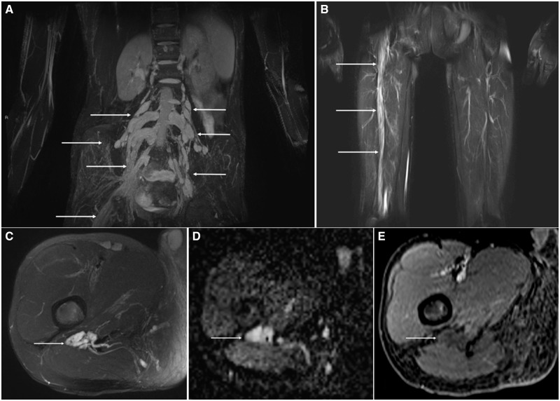
FIGURE 1.
Imaging features of localized hypertrophic neuropathy in RASopathy. Coronal Short tau inversion recovery (STIR) Sampling Perfection with Application optimized Contrasts using different flip angle Evolution (SPACE) maximum intensity projection (MIPs) through the pelvis (A) and thighs (B) shows multifocal, bilateral lumbar nerve root thickening (arrows) extending into the right lumbosacral plexus and sciatic nerve root (arrow). Axial T2 fat suppressed (FS) image through the proximal thigh (C) shows marked thickening of the sciatic nerve (arrow) without a discrete mass. Note, the absence of imaging features of a peripheral nerve tumor such as a target sign. Apparent diffusion coefficient map through the right thigh (D) also reveals marked thickening of the sciatic nerve (arrow) without a discrete mass or restricted diffusion to suggest a hypercellular neoplasm. Axial T1-FS post contrast image through right thigh (E) also reveals marked thickening of the sciatic nerve (arrow) without internal enhancement.

