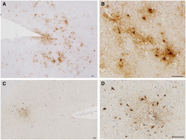
Figure 1.
Sulcal depth astrocytic and neuronal tau pathologies in CTE. (A and B) PHF1 immunohistochemistry reveals NFT and TSA pathology concentrated at the depths of cortical sulci of a former American football player (Case 3) and (C and D) chronic survivor of a single severe TBI (Case 11). (B and D) High magnification images from the same sulci showing perivascular pathology composed of mixed neuronal and astrocytic populations consistent with the preliminary diagnostic criteria for CTE. Scale bars = 100 µm.

