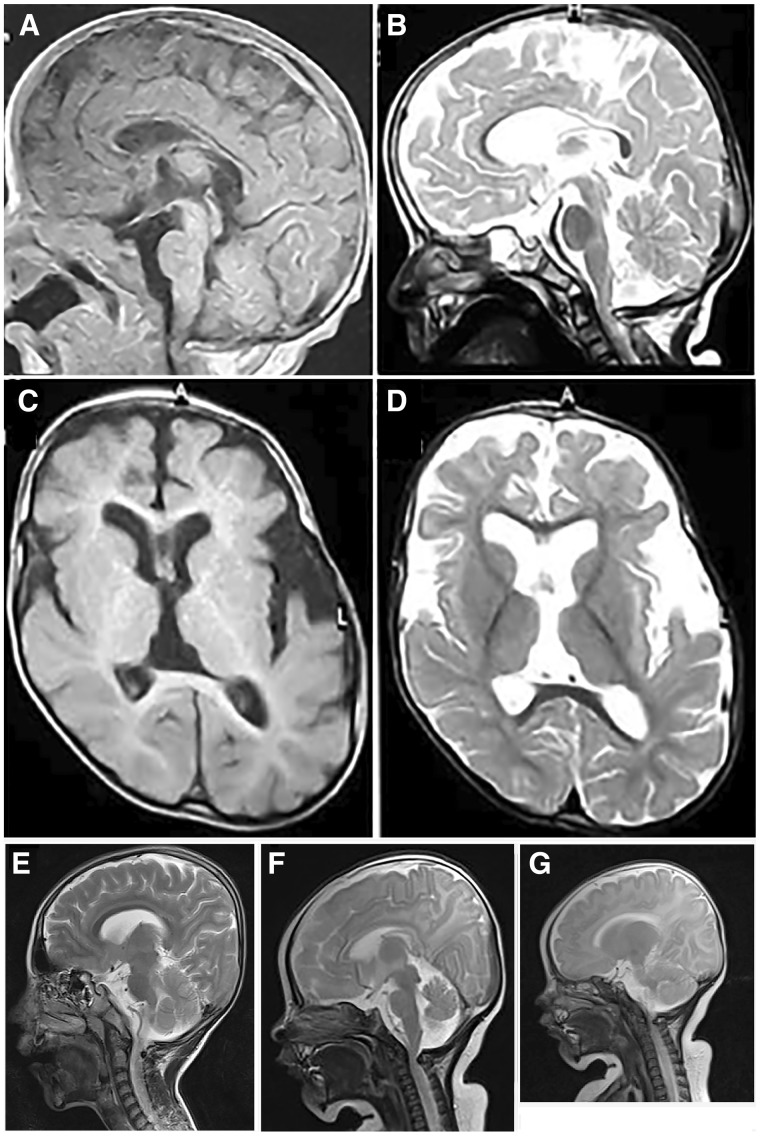
Figure 4.
Brain MRI. (A–D) Case 3 (Patient B-IV:4). Sagittal T1 and T2 cuts through the midline at the age of 1 month (A) and 2 years (B) showing hypoplastic corpus callosum, mainly body and to a lesser degree its genu, progressive cerebellar and cerebral atrophy and a cervical notch. Axial cuts T1 (C) and T2 (D) at the age of 2 years demonstrating mild-to-moderate cortical atrophy predominating on left hemisphere and mildly enlarged lateral ventricles. (E–G) Cases 7 and 8 (Patients D-V:2 and D-V:3). (E) Sagittal MRI image of Case 7 (Patient D-V:2) showing prominent inner liquor spaces without overt brain malformations. (F) Sagittal MRI image of Case 8 (Patient D-V:3) showing prominent inner liquor spaces without apparent brain malformations except a cervical notch causing an impression in the upper cervical spinal cord area. (G) Normal cerebellar size is demonstrated in Case 8 (Patient D-V: 3).

