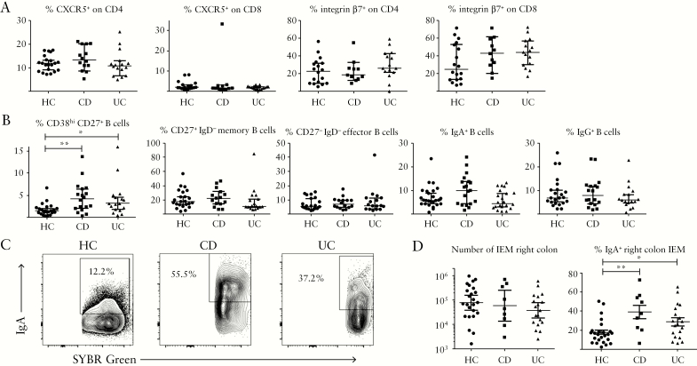
Figure 4.
Immunopathology of quiescent IBD reflects B cell dysregulation. A: Proportions of Tfh-like [CXCR5+] CD4/CD8 T cells and gut-homing [integrin-β7+] T cells in PBMC of healthy control and IBD donors. B: Circulating plasmablasts [CD38hi CD27+ CD19+] and B cell subsets in healthy and IBD donors. Kruskal-Wallis tests were used to compare groups [n = 23 HC; n = 18 CD; n = 17 UC]. C: IgA coating of IEM obtained from right colon biopsies of example HC, CD, and UC donors, after gating on SYBR Green+ events. D: Pooled data showing proportions of IgA+ IEM in donor groups. One-way ANOVA was used to compare groups [n = 25 HC; n = 9 CD; n = 19 UC]. IBD, inflammatory bowel disease; PBMC, peripheral blood mononuclear cells; HC, healthy controls; CD, Crohn’s disease; UC, ulcerative colitis; IEM, intraepithelial microbes.

