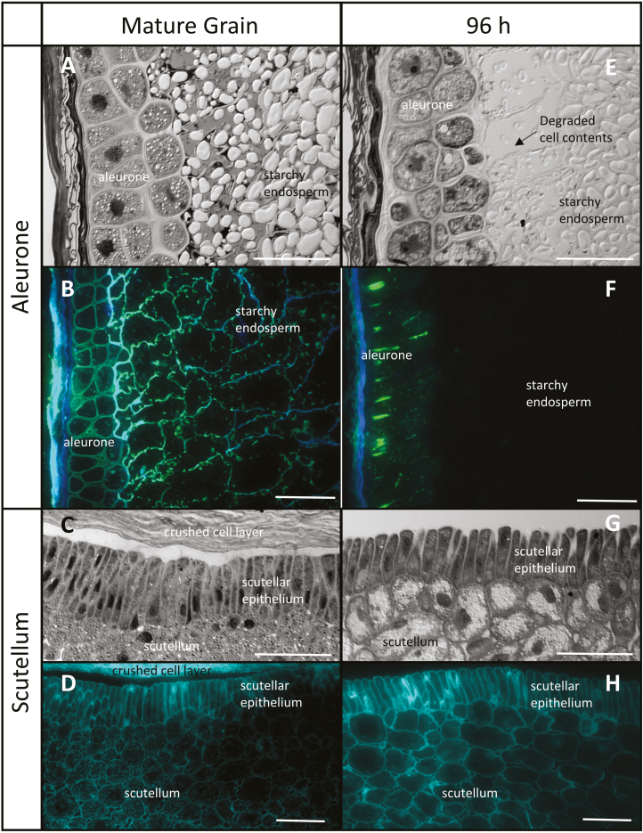
Fig. 1.
Comparative morphology of the aleurone layer and scutellum of mature and germinated barley grain. (A and E) Aleurone sections stained with toluidine blue and photographed using differential interference contrast (DIC) microscopy, showing the degradation of the subaleurone starchy endosperm between 0 h and 96 h. (B and F) Immunocytochemistry of aleurone arabinoxylan and (1,3;1,4)-β-glucans using LM11 (blue) and BG1 (green) antibodies. (C and G) Scutellum sections stained with toluidine blue and photographed using DIC microscopy, showing the lateral separation of the epithelial cells after 96 h. (D and H) Calcofluor staining of scutellum (turquoise). Scale bar=50 μm.

