Fig. 5.
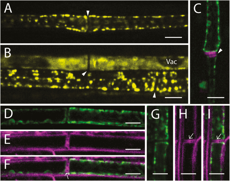
AtSuSy5–YFP and AtSuSy6–YFP were confined to the sieve elements of petioles. (A, B) Longitudinal sections of leaf petioles showing AtSuSy5 in mature sieve elements (A) and in immature sieve elements (B) containing vacuoles (Vac). (C) YFP labeling of AtSuSy6 (green) in mature sieve elements; the magenta signal indicates the sieve plates (white arrowhead) stained with aniline blue. (D–F) Longitudinal sections of leaf petioles showing the cytoplasmic localization of AtSuSy5. YFP signal of AtSuSy5 (green) is shown in (D), while the plasma membrane stained with FM4-64 (magenta) is shown in (E). Merged images of SuSy5–YFP and plasma membrane stained with FM4-64 are shown in (F). Plasmodesmata are indicated by an arrow. (G–I) Longitudinal sections of leaf petioles showing AtSuSy6–YFP (green) in (G) while the plasma membrane is labeled with FM4-64 (magenta) in (H). Merged images of SuSy6–YFP and plasma membrane stained with FM4-64 are shown in (I). Scale bars=5 μm.

