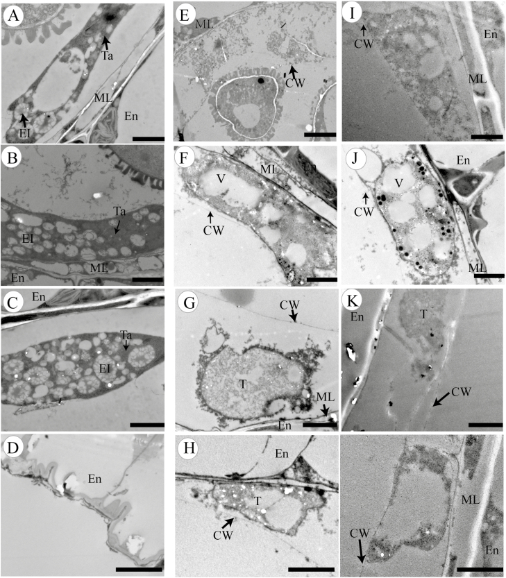
Fig. 5.
Transmission electron micrographs of anthers from the wild type and βvpe mutant
(A–D) Wild type; (E–H) βvpe mutant CS_1007412; (I–L) βvpe mutant SAIL_50_F12: (A), (E), and (I) stage 9; (B), (F), and (J) stage 10; (C), (G), and (K) stage 11; (D), (H), and (L) stage 12. Scale bar=2 μm. CW, cell wall; El, elaioplast; En, endothecium; ML, middle layer; T, tapetal cell; Ta, tapetosome; V, vacuole.

