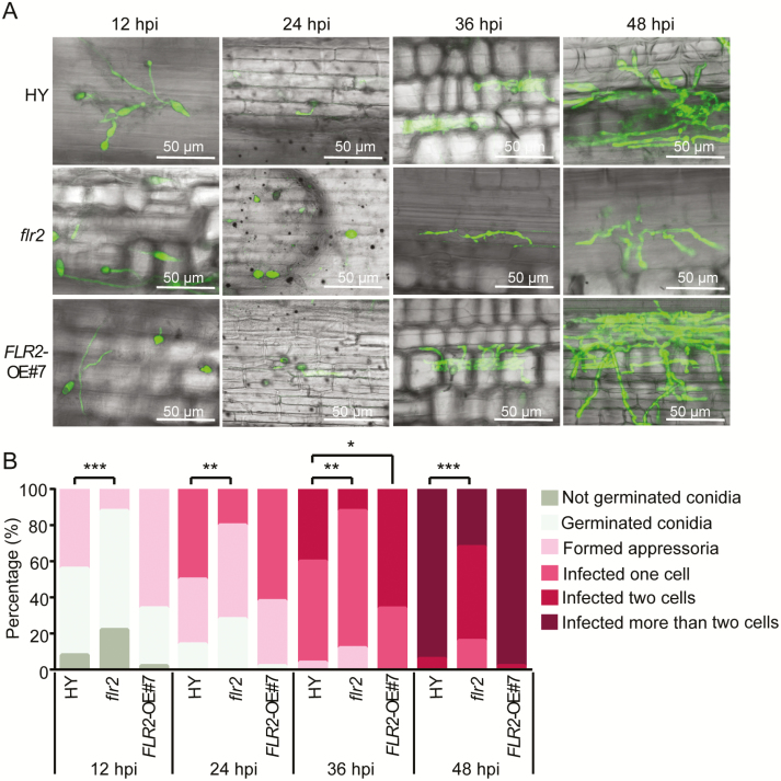
Fig. 5.
Fluorescence signals of GFP-tagged Magnaporthe oryzae strain 70-15GFP invasive hyphae in the leaf sheaths of flr2, FLR2-OE, and HY wild-type plants. (A) Confocal images showing the invasive M. oryzae hyphae in the epidermal cells of flr2, FLR2-OE#7, and HY leaf sheaths. Scale bar=50 μm. (B) Percentage of invasive hyphae growth at 12, 24, 36, and 48 hpi. The asterisks indicate significant differences from the control as determined by Crosstabs, followed by a χ 2 test (*P<0.05, **P<0.01, ***P<0.001). Fifty hyphae were counted per replication, and the experiment was repeated three times, each yielding similar results. (This figure is available in color at JXB online.)

