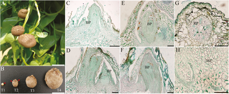
Fig. 1.
Morphology of the bulbil during key developmental stages. (A) Bulbil phenotype. (B) Photographs of the bulbil at the initiation (T1), early (T2), middle (T3), and mature stages (T4). (C–F) Paraffin sections of bulbils at T1 (C), T2 (D), T3 (E), and T4 (F) stages. The images show the zone of the junction region between the bulbil and axil. (G) Showing root primordia (RP). (H) Showing the meristematic zone (MZ). The arrows in T1 and T2 show the BP and root primordia, respectively. Scale bars (A and B), 1 cm; (C), 500 µm; (D–H), 200 µm.

