Fig. 2.

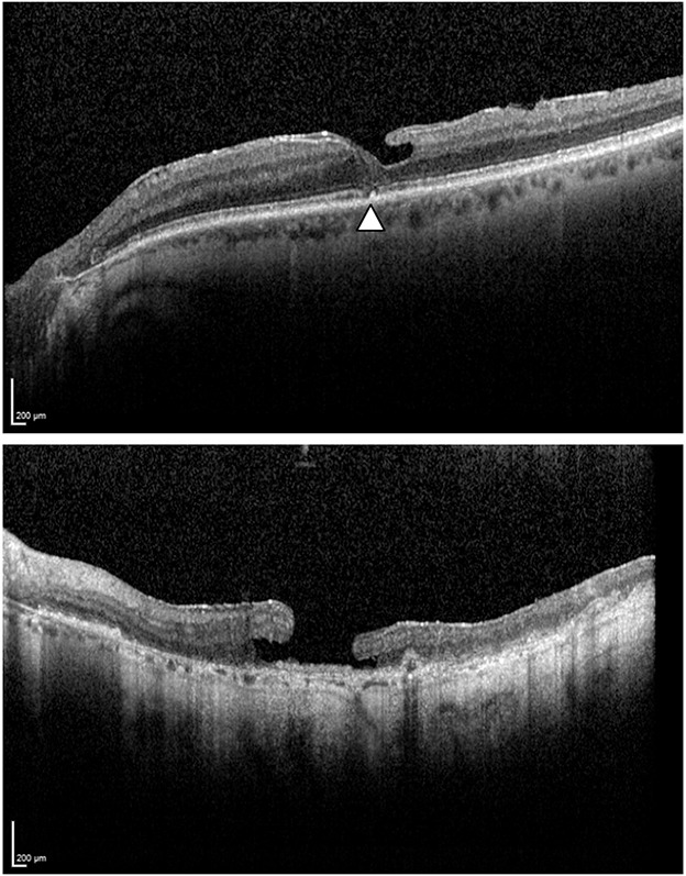
Ellipsoid zone integrity. Top: The arrowhead shows an ellipsoid zone disruption. Bottom: Ellipsoid zone absence with diffuse alteration in the outer retinal layer.

Ellipsoid zone integrity. Top: The arrowhead shows an ellipsoid zone disruption. Bottom: Ellipsoid zone absence with diffuse alteration in the outer retinal layer.