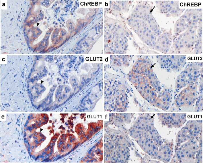
Fig. 5.
GLUT1 but not GLUT2 protein co-expressed in ChREBP-positive hepatocytes in HCC. Immunohistochemistry of ChREBP-positive malignant hepatocytes (a) showing GLUT2 negative (c) but GLUT1 positive (e) staining. Arrow heads indicating the same cell. Immunohistochemistry of ChREBP-negative malignant hepatocytes (b) showing GLUT2 positive on the membrane (d) but GLUT1 negative staining (f). Arrow indicating the same cell. Scale bar = 30 μm

