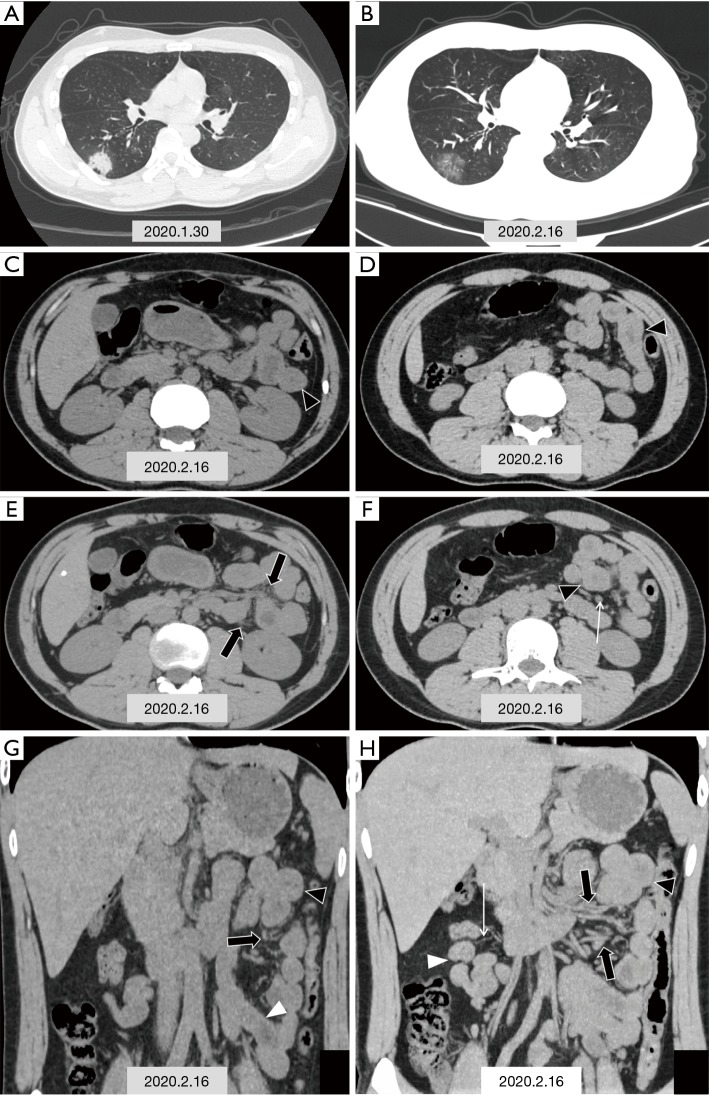
Figure 1.
CT images in a 29-year-old man with fever and diarrhea, and RT-PCR of nasopharyngeal swab and anal swab samples tested positive for SARS-CoV-2. (A) Chest CT scan obtained on the first admission on January 30th, 2020 shows one typical consolidative lesion among multiple opacities in subpleural region of the right lower lobe (black triangle); (B,C,D,E,F,G,H) chest and abdominal CT scans obtained on February 16th, 2020; (B) show the density of some patchy solid shadows reduced, demonstrating a ground glass density similar to “melting sugar” (black triangle); (C,D,E,F) axial images of abdominal CT scan show segmental wall swelling (black triangle) involving a segment of the jejunum in the left upper quadrant, with thickened mesenteric vessels (black arrow) and mild lymph nodes enlargement (white arrow); (G,H) coronal images of abdominal CT scan display segmental wall swelling of the jejunum (black triangle), with mesenteric vessels dilatation (black arrow). white triangle: normal intestinal wall, white arrow: mesenteric vessels.

