Figure 1.
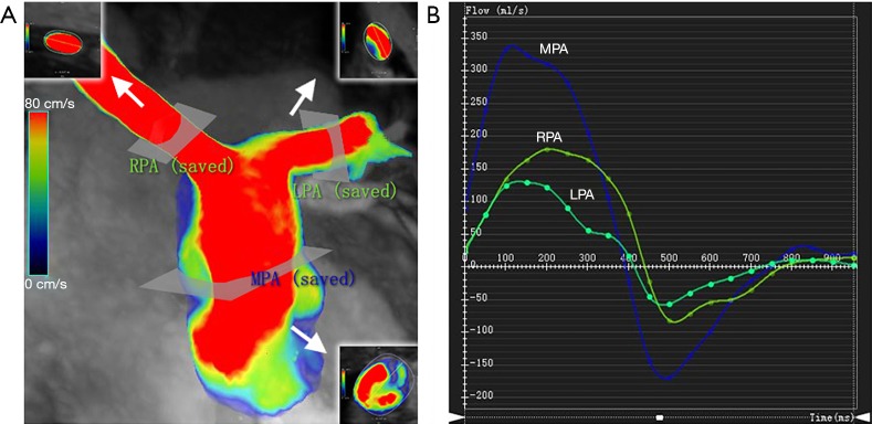
Routine blood flow parameters were analyzed in the pulmonary artery. (A) Locations of the MPA, RPA, and LPA cutplanes, which were placed approximately 10–15 mm downstream from the pulmonary valve or LPA/RPA bifurcation. (B) The results of peak velocity in MPA, RPA, and LPA cutplanes. MPA, main pulmonary artery; RPA, right pulmonary artery; LPA, left pulmonary artery. Arrows indicate sliced sheet of pulmonary artery.

