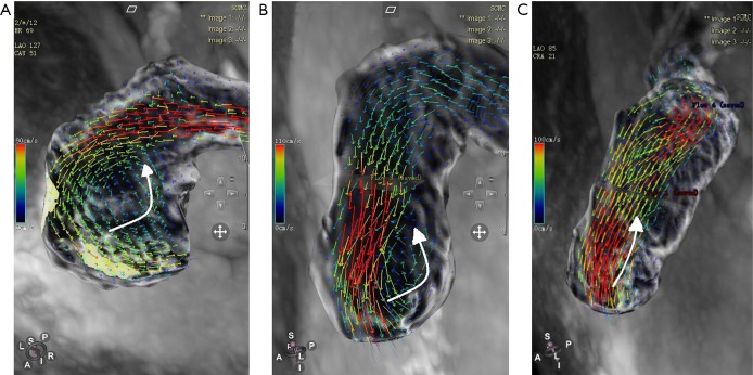
Figure 2.
Vortex severity was graded in the rTOF patients. (A) Follow-up magnetic resonance imaging of a 12-year-old, female child using a 4D flow sequence. During diastole, the flow pattern of an rTOF patient with severe PA dilation can be seen (group 1). A severe vortex formed in the PA, and the flow velocities were lower; the vortex formed by regurgitation was close to the pulmonary valve. (B) Follow-up magnetic resonance imaging of a 9-year-old, male child using 4D flow sequence. During diastole, a flow pattern of an rTOF patient with PA was observed, and the vortices formed in the PA and mild vortex locally affected the wall shear stress inside the vessel (group 2). A vortex formed by regurgitation found close to the pulmonary valve. (C) Follow-up magnetic resonance imaging of a 15-year-old, male teenager using 4D flow sequence. During diastole, none-flow grading appeared in the pulmonary artery (group 3). rTOF, repaired tetralogy of Fallot; PA, pulmonary artery. Curved arrows indicate direction of blood flow.

