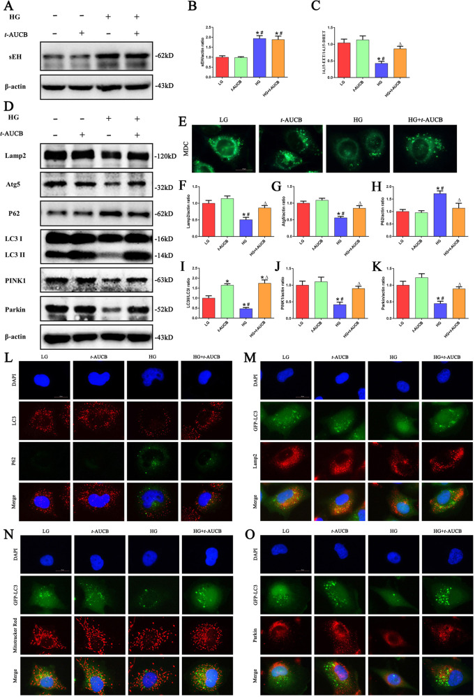
Fig. 3. Inhibition of sEH restored HG -induced impaired autophagy flux and mitophagy.
a Western blot analysis of sEH in HK-2 cells treated with HG or/and t-AUCB for 24 h. b Densitometric analysis of sEH (n = 3, *P < 0.05 vs. LG, #P < 0.05 vs. t-AUCB). c ELISA kit analysis of sEH enzymatic activity (14,15-EET to 14,15-DHET ratios) in HK-2 cells (n = 3, *P < 0.05 vs. LG, #P < 0.05 vs. t-AUCB, ΔP < 0.05 vs. HG). d Western blot analysis of Lamp2, Atg5, LC3-II/LC3-I, p62, PINK1 and Parkin in HK-2 cells treated with HG or/and t-AUCB for 24 h. e Representative images of MDC staining in different groups of HK-2 cells after treated with HG or/and t-AUCB for 24 h. f–k Densitometric analysis of Lamp2, Atg5,LC3-II/LC3-I, p62, PINK1 and Parkin in figure d (n = 3, *P < 0.05 vs. LG, #P < 0.05 vs. t-AUCB, ΔP < 0.05 vs. HG). l Representative images of immunofluorescence double labeling of LC3 and P62 in different groups of HK-2 cells after treated with HG or/and t-AUCB for 24 h. m–o Colocalization analysis of immunofluorescence images of GFP-LC3 (green) and Lamp2 (red), GFP-LC3 (green) and Mito Tracker (red), GFP-LC3 (green) and Parkin (red) in different groups of HK-2 cells after treated with HG or/and t-AUCB for 24 h.

