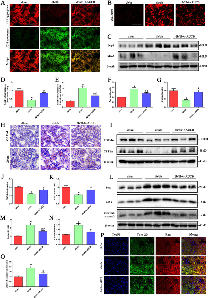
Fig. 7. t-AUCB administration attenuated mitochondrial dysfunction, ROS production, and apoptosis in tubules of db/db mice.
a Kidney sections stained with JC-1. b Kidney sections stained with Mito SOX. c Western blot analysis of Drp1and Mfn2 expression in kidney tissues. d Quantification of the fluorescence intensity of JC-1 staining in figure a. e Quantification of the fluorescence intensity of Mito SOX staining in figure b. f, g Densitometric analysis of Drp1and Mfn2 expression in the figure c. h Kidney sections stained with Oil Red O staining. i Western blot analysis of PGC-1α and CPT1A expression in kidney tissues. j, k Densitometric analysis of PGC-1α and CPT1A expression in figure i. l Western blot analysis of Bax, Cyt c and cleaved-caspase3 expression in kidney tissues. m–o Densitometric analysis of Bax, Cyt c and cleaved-caspase3 expression in the figure l. p Representative images of immunofluorescence double labeling of Tom20 and Bax in different kidney tissues. *P < 0.05 vs. db/m, #P < 0.05 vs. db/db.

