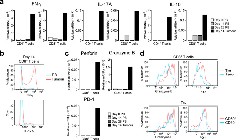
Figure 3.
CD8+ TILs express high levels of cytotoxic molecules and PD-1. (a) Relative mRNA expression of IFN-γ, IL-17A and IL-10 in CD4+ and CD8+ T cells isolated from PB and tumour tissue. Blood was obtained at days 0, 14 and 28, and the tumour was resected at day 14 after tumour transplantation. The mRNA levels were assessed by real-time PCR and are presented relative to β-actin levels. (b) Intracellular staining for IFN-γ and IL-17A in CD8+ T cells from PB and tumours. Blood and the tumour were obtained at day 14. (c) Relative mRNA expression of perforin, granzyme B and PD-1 in CD8+ T cells isolated from PB and tumour tissue. The mRNA levels were assessed as in (a). (d) Flow cytometric analysis of the expression of granzyme B and PD-1 in CD8+ T cells and CD8+ TEM cells from tumours.

