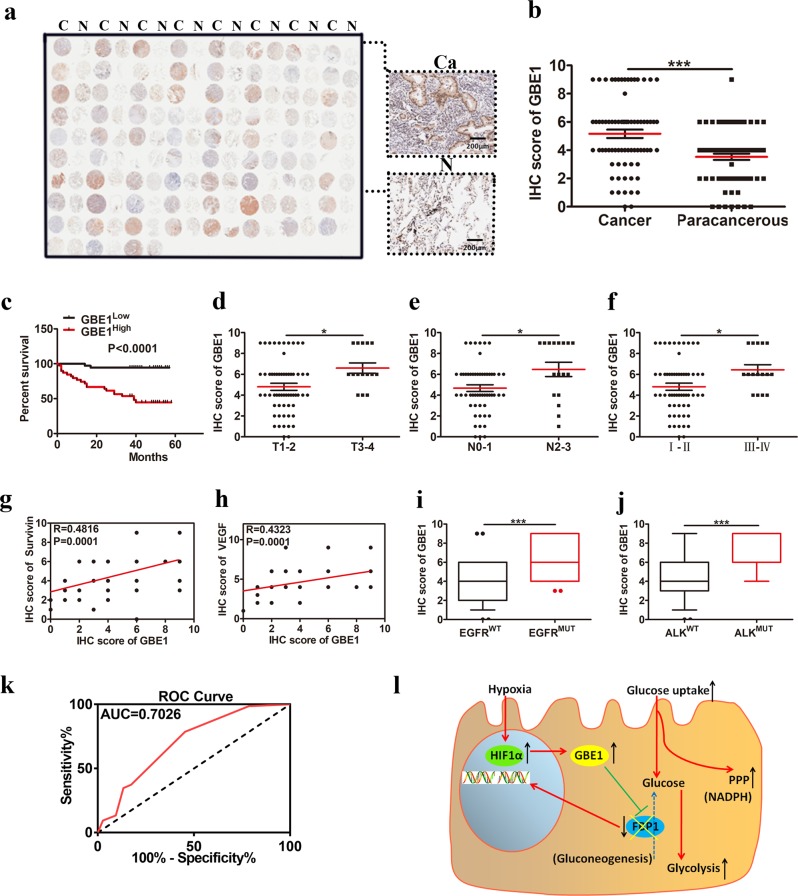
Fig. 7.
GBE1 is a negative prognostic biomarker for LUAD patients. a IHC staining of GBE1 expression from a representative human LUAD tissue microarray. C cancer tissue, N adjacent normal lung. b Quantification of the IHC staining showing GBE1 expression in 75 LUAD and paired normal lung tissues. c Kaplan−Meier OS curve based on high or low GBE1 expression. d–f Scatter plot of GBE1 expression based on IHC score in the LUAD tissues with primary tumors (d) in regional lymph nodes (e) and by tumor stage (f). g, h Correlation between survivin and VEGF expression with GBE1 in the LUAD tissues. i, j IHC score of GBE1 in the LUAD tissues according to EGFR or ALK mutation status. k ROC curve based on GBE1 expression in the LUAD tissues. l Graphical summary of the metabolic pathway of GBE1 under hypoxia in the LUAD cells. Data are represented as the means ± SD. *P < 0.05, ***P < 0.001

