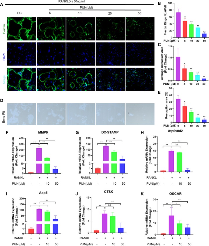
Figure 2.
PUN inhibited F-actin rings formation, bone-resorbing activity, and osteoclast-specific genes expression. (A) After treatment with or without PUN, RAW 264.7 macrophages were incubated with induction medium for 4 days and then stained for F-actin. F-actin (green) and nuclei (blue) staining of osteoclasts were photographed at the magnification of 320x by fluorescence microscope. (B) Quantification of the F-actin ring number per well. (C) Quantification of the relative area of osteoclast. (D) RAW 264.7 macrophages were plated on bone resorption plates and cultured with induction medium containing 50 ng/ml RANKL together with various concentrations of PUN (0, 5, 10, 20, and 50 μM) for 4 days. Resorption pits were visualized at the magnification of 320× under an inverted microscope. (E) Quantification of pits formation area. All bar graphs are presented as mean ± SD. *p < 0.05, **p < 0.01 compared with control group, n = 3 per group. (F–K) RAW 264.7 macrophages were incubated in six-well plate in triplicate with induction medium containing 50 ng/ml RANKL and various concentration of PUN for 24 h. The gene copies of MMP9, DC-STAMP, Atp6v0d2, Acp5, CTSK, and OSCAR were quantified by RT-PCR. *p < 0.05, **p < 0.01 compared with RANKL group, n = 6 per group. NS, not statistically significant.

