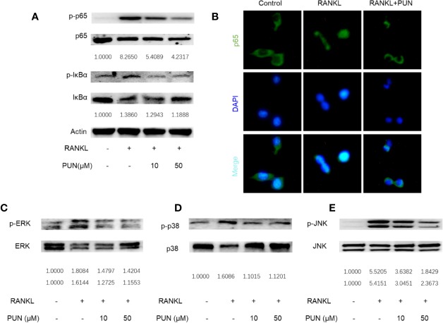
Figure 3.
PUN interfered with RANKL-induced activation of NF-κB and MAPKs pathways. After presentative treatment with or without various concentration of PUN (0, 10, and 50 μM) for 6 h, RAW264.7 cells were stimulated by 50 ng/ml RANKL. Then, the cells were collected and lysed for Western blot analysis (A, C–E). The relative grey level of phosphorylated protein of p65, IκBα, p-ERK/MAPK, p-38/MAPK, and JNK to total protein were quantified by using Image Lab software and compared with control group. (B) After treated with or without PUN (50 μM), RAW264.7 cells were stimulated by 50 ng/ml RANKL for 1 h and then stained for p65 antibody and secondary antibody with FITC. And the p65 nuclear localization was visualized using an immunofluorescence microscope. n = 3 per group.

