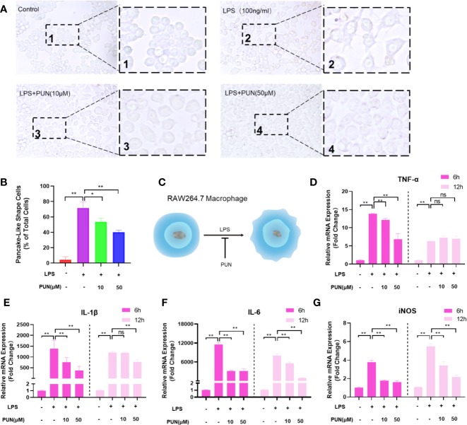
Figure 6.
PUN suppressed LPS induced inflammation. (A) After presentative treatment with or without various concentration of PUN (0, 10, 50 μM) for 6 h, RAW264.7 cells were stimulated by 100 ng/ml LPS for 24 h. Pancake-like shape cells were visualized at the magnification of 320× under an inverted microscope. (B) Quantification of the percentage of pancake-like shape cells. (C) The schematic diagram of the effect of PUN on the cellular shape of RAW 264.7 macrophages. (D–G) After treated with or without PUN, RAW 264.7 macrophages were incubated in six-well plate with 100 ng/ml LPS and various concentration of PUN for 6 or 12 h. The pro-inflammatory cytokines gene copies of TNF-α, IL-1β, IL-6, and iNOS were quantified by RT-PCR. *p < 0.05, **p < 0.01 compared with LPS group, n = 6 per group. NS, not statistically significant.

