Figure 3.
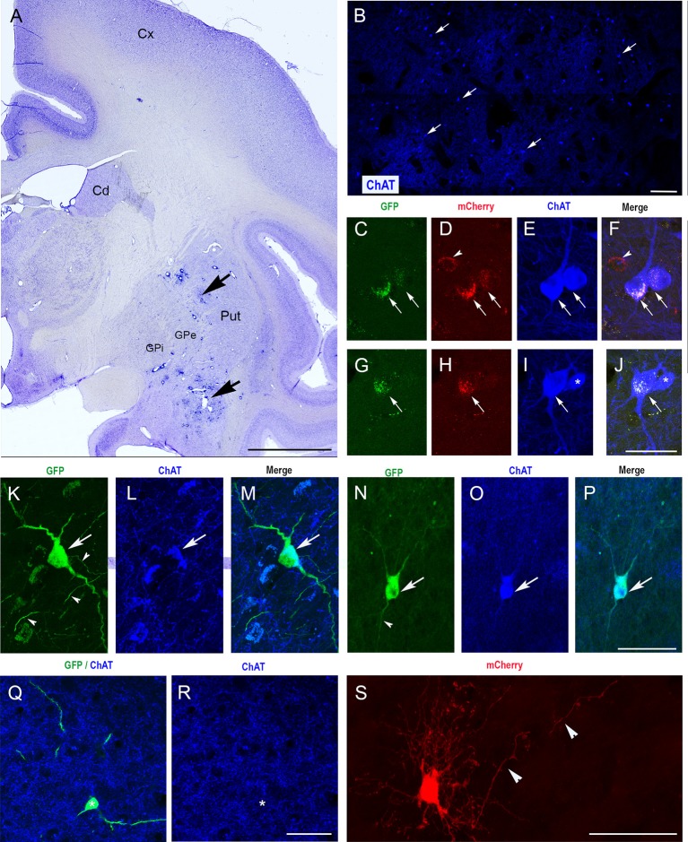
Selective expression of GFP in ChIs of the monkey striatum with CAV-2 containing the human ChAT promoter (CAV-pChAT-GFP). (A) Coronal section of a macaque brain stained with Cresyl violet showing the injection sites of CAV-pChAT-GFP in the striatum. (C–S) Immunofluorescence labeling for simultaneous detection of, GFP (green), mCherry (red) and ChAT (blue) in an adjacent coronal section. Images of the fluorophores obtained by sequential acquisition of separate wavelength channels from a single confocal slice. (B) Distributions of ChAT-containing neurons within the striatum. (C–J) In monkey C in which both CAV-2 vectors were injected in the same hemisphere, GFP and mCherry expression were restricted to the cell bodies. Most of these neurons that were labeled for both GFP (green, arrows) and mCherry (red, arrows) contained ChAT (blue) whereas neurons expressing mCherry only (red, arrowheads) were not labeled for ChAT. (K–S) In Monkey P in which CAV-pChAT-GFP and CAV-mCherry were injected in the right and left striatum, respectively most neurons expressing GFP (K,N,Q, green) or mCherry (S) displayed well-labeled cell bodies as well as dendritic and axonal (arrowheads) processes. (K–P) Most neurons expressing GFP (green, arrows) contained ChAT (Blue, arrows). (Q,R) A few neurons expressing GFP did not show detectable levels of ChAT. (F) Merge of (C–E). (J) Merge of (G–I). (M) Merge of (K,L). (P) Merge of (N,O). Scale bars: (A), 5 mm; (B), 600 μm; (C–S) 50 μm.

