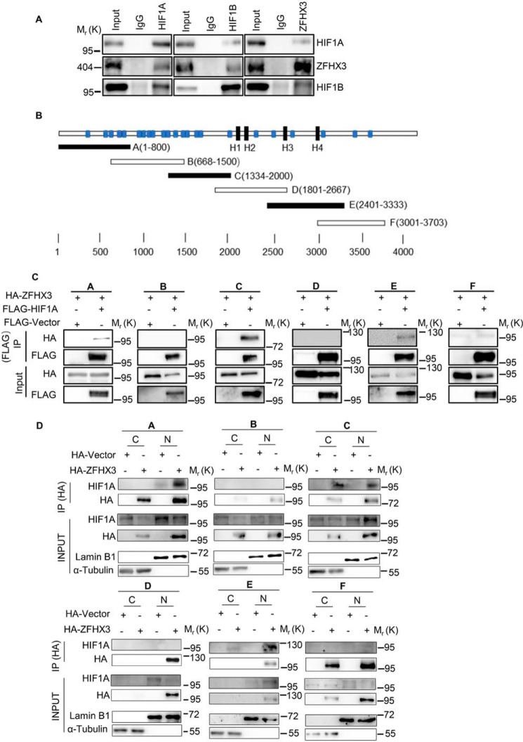
Figure 5.
ZFHX3 physically interacts with the HIF1A complex. A, detection of protein association between HIF1A, ZFHX3, and HIF1B in HepG2 cells by co-IP with HIF1A (left), HIF1B (middle), or ZFHX3 antibody (right) and Western blotting with the indicated antibodies. Input (1/20 of whole-cell lysate) indicates cell lysate not subjected to IP. B, schematic of full-length ZFHX3 (3703 residues, horizontal bar) with four homeodomains (black rectangle) and 23 zinc fingers (blue rectangle). The six overlapping fragments of ZFHX3 were named A–F. C, six HA-tagged overlapping ZFHX3 fragments and FLAG-tagged HIF1A were ectopically expressed in HepG2 cells under hypoxia, and IP and Western blotting were applied to test ZFHX3–HIF1A interactions. D, association of HIF1A with different fragments of ZFHX3 in HeLa cells ectopically-expressed HA-tagged ZFHX3 fragments under hypoxia. Nuclear and cytoplasmic fractions were separated, and IP and Western blotting were applied to each fraction. Input (1/50 of cytoplasmic or nuclear lysate) indicates the lysate not subjected to IP. C, cytoplasm; N, nucleus.

