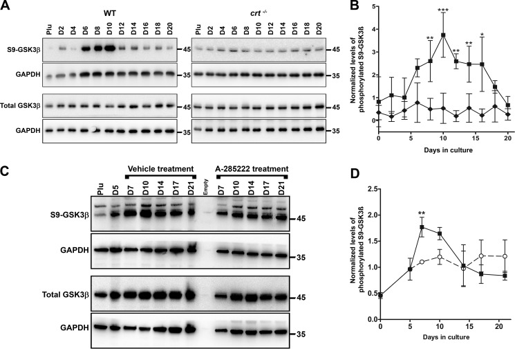
Figure 5.
Calreticulin and NFAT regulate GSK3β-deactivation. A and C, Western blot analysis of whole-cell lysates harvested at the indicated times from WT control, crt−/− ESCs (A), or WT ESCs treated with A-285222 for days 6–17 undergoing osteoblast differentiation (C). Utilized antibodies were specific to GSK3β, serine 9–phosphorylated GSK3β, and GAPDH as an internal control. B and D, quantitative representation of the density of the Western blotting bands of phosphorylated GSK3β normalized to internal control GAPDH. Data are expressed as means ± S.D. (error bars), n ≥ 3. Shown are WT (■), crt−/− (♦) ESCs, and WT ESCs treated with A-285222 (○); two-way ANOVA. B, p < 0.0001, F = 80.51. D, p = 0.018, F = 6.448. Bonferroni post hoc analysis was as indicated: *, p < 0.05; **, p < 0.01; ***, p < 0.001.

