Figure 6.
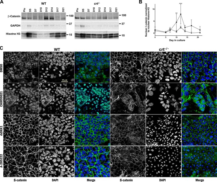
Calreticulin regulates levels of nuclear β-catenin during osteoblast differentiation. A, Western blot analysis of nuclear fractions extracted from WT control and crt−/− ESCs on the indicated days. B, quantitative representation of the density of the Western blotting bands (WT control (■) and crt−/− ESCs (♦)). Utilized antibodies were specific to β-catenin and the internal nuclear and cytosolic standards, histone H3 and GAPDH, respectively. Data are expressed as means ± S.D. (error bars), n ≥ 3; two-way ANOVA of band densities: B, p < 0.0001 and F = 19.76. Bonferroni post hoc test was as indicated: **, p < 0.01; ***, p < 0.001. C, confocal immunofluorescence images of localization of β-catenin at day 14 of the osteogenic differentiation protocol of WT control ESCs or crt−/− ESCs with or without treatment starting at day 6 by the molecules labeled on the left. Dual-channel greyscale images of a single field are displayed with β-catenin in the left panel, DAPI-stained nuclei in the right panel, and the RGB panel of a merged image of green (β-catenin) and blue (DAPI). Scale bar, 10 μm.

