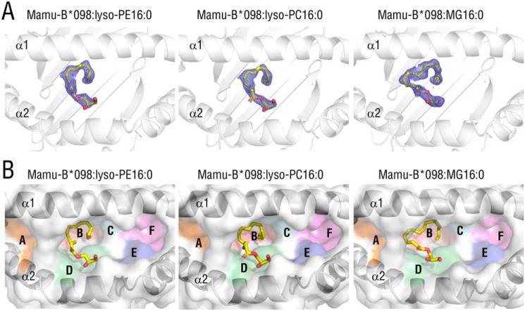
Figure 2.
Crystal structures of Mamu-B*098 complexed with lyso-PE16:0, lyso-PC16:0, and MG16:0. A, top views of the Ag-binding groove of Mamu-B*098 complexed with lyso-PE16:0 (left), lyso-PC16:0 (middle), and MG16:0 (right) are displayed with ribbon models of heavy chains and 2Fo − Fc maps (contoured at 0.8σ) for bound ligands (yellow sticks). The maps obtained for bound lysophospholipids only correspond to the structure of monopalmitoylglycerol, and, thus, the headgroup structures are not included. B, the surface of the antigen-binding groove is shown with pockets A–F colored, and the bound ligands are also displayed as yellow sticks.

