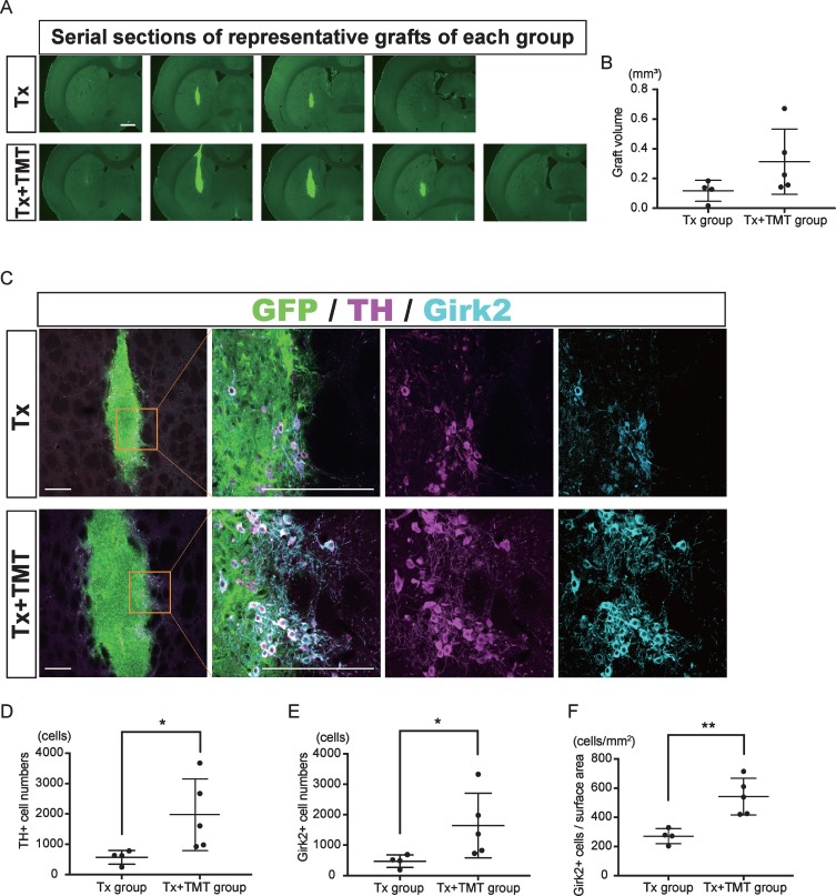
Fig.2.
Exercise increased the number of dopamine neurons in the graft. (A) Serial sections of representative grafts at 6 weeks after transplantation in Tx and Tx+TMT rats. The grafts were identified with the expression of GFP. (B) Quantification of the graft volume from the Tx group (N = 4) and Tx+TMT group (N = 5). (C) Immunofluorescence images of the grafts. (D, E and F) Quantification of TH+ (D) and Girk2+ (E, F) cells in the grafts. The number of TH-positive cells was significantly larger in the Tx+TMT group (D). More Girk2-positive cells survived in the Tx+TMT group, both in total number (E) and in number adjusted by the surface area of the graft (cells/cm2, F). *p = 0.016 by Mann-Whitney test, **p = 0.005 by Unpaired t test. All values are expressed as the mean±SD (B, D, E and F). Scale bars: 1 mm (A), 200 μm (C).

