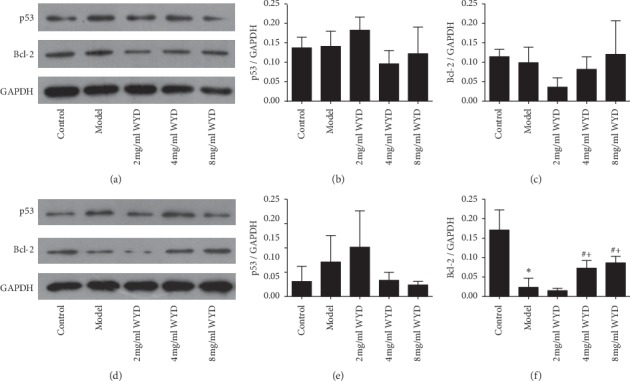
Figure 6.

Effects of WYD on p53 and Bcl-2 protein expression in the MN cell model. Podocytes were incubated with RPMI-1640 medium containing serum from volunteers or serum from patients and different concentrations of WYD for 24 h (a, b, and c) and 48 h (d, e, and f). (a and d) The protein levels of p53 and Bcl-2 were analyzed using Western blot. For densitometry, GAPDH was used as an internal control. A reduction in p53 expression and an increase in Bcl-2 expression were detected in the 4 mg/ml and 8 mg/ml WYD groups compared with the model group both for 24 h and 48 h incubation. (b, c, e, and f) Bar graphs show the expression of p53 and Bcl-2. WYD, Wenyang Lishui decoction. ∗P < 0.05 vs. the control group. #P < 0.05 vs. the model group. +P < 0.05 vs. the 2 mg/ml WYD group.
