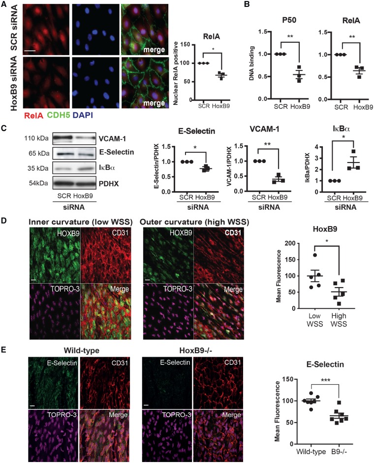
Figure 3.
HoxB9 promotes inflammatory activation in response to low WSS. (A–C) HUVECs were transfected with siRNA targeting HoxB9 or with scrambled control sequences. Cells were then exposed for 72 h to low (centre) WSS using the orbital system. (A) Nuclear expression of RelA was assessed by immunostaining (in red) in combination with stainings for nuclei (DAPI-blue) and endothelial marker CDH5 (green). Bar = 10 μm. (B) The activity of RelA and p50 NF-κB subunits was assessed by DNA-binding ELISA. Mean optical density (OD) 450 nm values ± SEM are shown. (C) The expression levels of VCAM-1 and IκBα were assessed by Western blotting using specific antibodies and anti-Calnexin was used to control for total protein levels. (D) The expression of HoxB9 was quantified at the inner curvature (low WSS) and outer curvature (high WSS) of the murine aortic arch by en face staining (green). N = 5. Bar = 10 μm. (E) Wild-type or HoxB9−/− mice were studied (N = 7/group). The expression of E-selectin was quantified at the inner curvature of the murine aortic arch by en face staining. Bar = 10 μm. (D and E) EC were identified using anti-CD31 antibodies (red) and nuclei were co-stained using TOPRO3 (purple). Differences between means were analysed using an unpaired (A, B, C, and E) or paired (D) t-test. *P < 0.05, **P < 0.01, ***P < 0.001.

