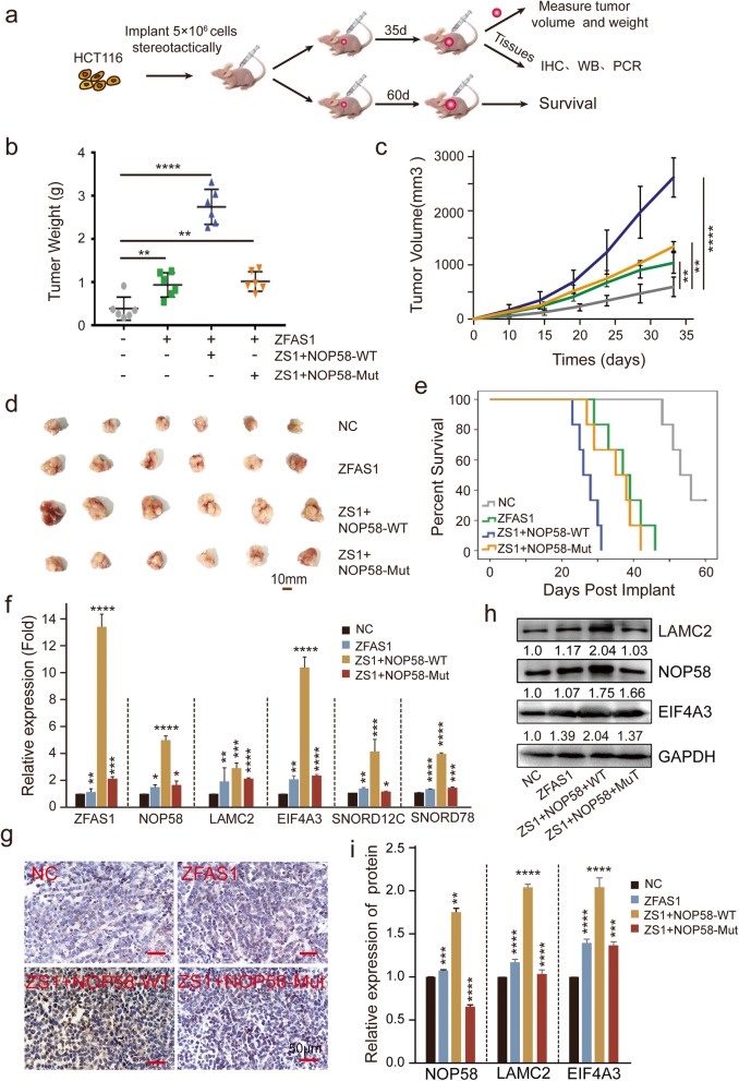
Fig. 7.
ZFAS1 inhibited proliferation by targeting NOP58 protein in vivo. a, Schematic diagram of xenografts in BALB/c nude mice by inoculating HCT116 cells that were stably co-transfected with ZFAS1, ZFAS1-NOP58-Wild, and ZFAS1-NOP58-Mut, as well as the control with empty vector at their right armpits. Then half of the xenografts were sacrificed at the 35th day after injection and the other half were tracked until death. b, Mean tumor weight of each group xenografts in nude mice. Data are shown as mean ± s.d., n = 6 for each group. c, Mean tumor volumes on different days for each group xenografts in nude mice. Data are showed as mean ± s.d., n = 6 for each group. d, Representative tumors size excised on day 35 are shown. e, Kaplan-Meier graph showing overall survival of each group, n = 6. f, qPCR assays were performed to determine the (m) RNA expression of ZFAS1, NOP58, LAMC2, EIF4A3, SNORD12C, SNORD78 in above each group. g, h and i, IHC assay and western blot to detect the protein expression of NOP58 in xenografts tumor tissues of each group. The groups were as follows: NC (empty vector); ZFAS1 (pcDH-ZFAS1); ZFAS1 + NOP58-Wild (co-transfected with pcDH-ZFAS1 and pcDH-NOP58-Wild); ZFAS1 + NOP58-Mut (co-transfected with pcDH-ZFAS1 and pcDH-NOP58-Mut).*, P < 0.05; **, P < 0.01; ***, P < 0.001; ****, P < 0.0001

