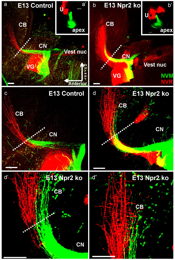
Fig. 2. Cerebellar afferent projections studied in 3 Npr2 mutants.

E13.5 Npr2 mutants (b,d) showed more vestibular (NCR, red) and cochlear (NVM, green) afferents in compared to littermate control animals (a,c) after comparable applications of dyes to the cochlea and vestibular organs (a’,b’). Higher power shows that many, but not all cochlear afferents go past the r1/2 border (indicated by dotted lines) in Npr2 mutants (d,d’,d”) but not in control animals (a,c). Cochlear afferents project at this stage almost as far medial as vestibular afferents (d”). CB, cerebellum; CN, cochlear nucleus. Red blood cells (green dots) are highly excited by the 635nm used for NVM. Orientation shown in (a) is identical for all images with dorsal up and anterior to the left. Bar indicates 100 μm.
