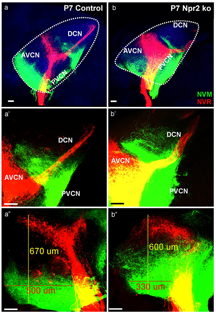
Fig. 8. Detailed analysis of two P7 Npr2 mutant (b,b’b”) and control littermate (a,a’a”) show overall cochleotopic matching projections with apical fibers (NVM, green) projecting ventral and basal fibers (NVR, red) projecting dorsal in the antero-ventral cochlear nucleus (AVCN).

However, while fibers in control animals remain restricted to narrow bands, in particular cochlear afferents from the apex expand to reach almost the basal turn projection in the mutant (compare a”,b”). Projection of the basal turn reaches unbranched to the dorsal cochlear nucleus (DCN; b’) whereas basal turn neurons show a profound collateral expansion along the ventral border of the DCN with the PVCN which is comparable in Npr2 mutant (b,b’) and control littermates (a,a’). In addition to the AVCN dorsal expansion, the anterior expansion of the basal turn is truncated to about 2/3 of the rostral extend (in the presented case from 500 um to 300 um). Note that branches emerge from the apical fibers to nearly fill the gap between apical and basal tonotopic tracts (a”, b”). Blue indicates the 488 background fluorescence that was used to outline with a dotted line the cochlear nuclei complex. Bar indicates 100 μm.
