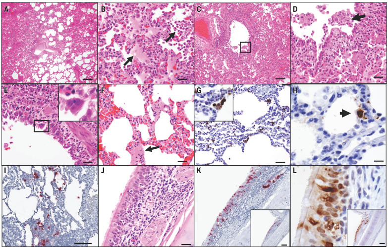
Fig. 3. SARS-CoV-2 induces acute viral interstitial pneumonia.
(A to F) Hematoxylin and eosin–stained sections of fixed lung tissue from SARS-CoV-2–infected rhesus macaques 2 days after challenge showing interstitial edema and regional lung consolidation (A), intra-alveolar edema and infiltrates of neutrophils (B), bronchiolar epithelial sloughing and necrosis [(C) and (D)], bronchiolar epithelial syncytial cell formation (E), and hyaline membranes within alveolar septa (F). (G and H) Immunohistochemistry for SARS-N showing virus-infected cells within interstitial spaces, including a viral syncytial cell within the lumen (G) and virus-infected alveolar lining cells (H). (I) Inflammatory infiltrate showing multiple cells containing SARS-CoV-2 RNA by RNAscope in situ hybridization. (J to L) Bronchial respiratory epithelium showing inflammation within the submucosa and transmigration of inflammatory cells into the ciliated columnar respiratory epithelium of a bronchus (J), SARS-CoV-2 RNA (K), and SARS-N (L). Scale bars: (A), 200 μm; (C), (I), (K), and (L), 100 μm; (G), 50 μm; (B), (D), (E), (F), and (J), 20 μm; (H), 10 μm.

