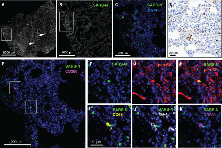
Fig. 4. SARS-CoV-2 infects alveolar epithelial cells in rhesus macaques.
Shown is CyCIF staining of fixed lung tissue from SARS-CoV-2–infected rhesus macaques 2 days after challenge. (A) Whole-slide image of a lung stained with Hoechst 33342 to visualize cell nuclei (grayscale); regions of nuclear consolidation (arrows) and foci of viral replication (box) are highlighted. (B) Higher-magnification image of inset box in (A) showing staining for SARS-N (green) and cell nuclei (grayscale). (C) Higher-magnification image of inset box in (B) showing SARS-N (green) and cell nuclei (blue). (D) Bright-field immunohistochemistry for SARS-N from corresponding lung region depicted in (C). (E to K) CyCIF staining for DNA (all panels, blue) and SARS-N [(E), (F), and (H) to (K), green], CD206 [(E) and (K), magenta], pan-CK [(G) and (H), red], CD68 [(I), yellow], or Iba-1 [(J), grayscale] showing virus-infected epithelial cells and macrophages near an infected epithelial cell. Scale bar for (F) to (K), 50 μm.

