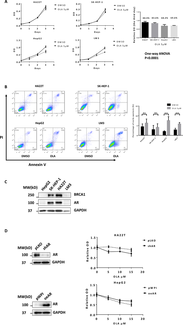
Figure 1.
Differential sensitivity of HCC cells to Olaparib treatment. A) Human HCC cell lines HA22T, SK-HEP-1, HepG2, and LM3 were treated with DMSO or 5 μM Olaparib. Cell viability was detected using MTT assay. B) PI/Annexin V apoptosis assay of HA22T, SK-HEP-1, HepG2, and LM3 by flow cytometry after 48h DMSO or 5 μM Olaparib treatment. C) Western blots showed protein expression levels of BRCA1 and AR in different HCC cell lines. D) HA22T and HepG2 cells were stably infected with lentiviruses expressing shRNA targeting AR or AR cDNA for overexpression AR. Western blots (left) showed the expression of AR and GAPDH (loading control). After manipulating AR, cells were treated with different concentrations of Olaparib for 2 days. The MTT assay was performed to determine cell viability (right). For A and B, quantitation are at the right. Data are presented as mean ± SD. **P < 0.01, NS=Not Significant.

