
Figure 2.
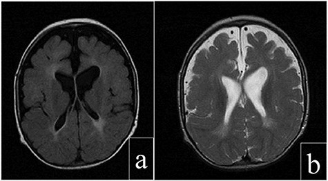
Cerebral magnetic resonance images of the patient: (a) axial T2 FLAIR view showing periventricular increased signal. (b) Axial FRFSE T2 view showing enlargement of the subarachnoid space, ventricles and sylvian fissures.
Official websites use .gov
A
.gov website belongs to an official
government organization in the United States.
Secure .gov websites use HTTPS
A lock (
) or https:// means you've safely
connected to the .gov website. Share sensitive
information only on official, secure websites.
Cerebral magnetic resonance images of the patient: (a) axial T2 FLAIR view showing periventricular increased signal. (b) Axial FRFSE T2 view showing enlargement of the subarachnoid space, ventricles and sylvian fissures.